


Cranial-Sacral Therapy (CST)
The cranial-sacral system addresses bones and membranes that surround the central nervous system. Restrictions here can strain neural tissues and affect coorespondingly innervated somatic areas, organs, or tissues. CST uses light manual techniques aimed at inducing relaxation responses that decrease, unwind, or remove neural tissue strain. CST, while particularly effective for treatment-resistant headaches, also have positive bearing on varied cases of unresolved chronic muscle and joint pain. Case studies show usefulness in helping resolve some digestive and neuro-endocrine disorders. Learning curves, attention spans, and memory retention are often reported improved.
Movement Therapies and Postural Re-alignment
Postural habituation and any form of trauma or sudden mal-positioning are the most common causes of tissue structure / function imbalance. Compensations include often inappropriate tissue lengthening and shortening. Somatic dysfunctions over time build up and bear heavily on joints and their surrounding / supporting structures. Muscle length / strength imbalance identification allows for both prevention and proper resolution of Neuro-myofascial syndromes. Postural and Movement Rehabilitation addresses these imbalances.
Lymphatic Drainage Therapy (LDT)
Trauma, physical deconditioning, lack of exercise, myofascial restrictions, and local tissue / area inflammation can cause lymph stagnation. Buildup of cellular debris and inflammatory mediators reinforce the nociceptive cycle and somatic dysfunctions. LDT can improves circulation and helps resolve edema. Joint and soft tissue swelling can be significantly reduced. Chronic pain syndromes can be improved or partially resolved.
Neuromuscular Techniques (NMT) / Neuro-myofascial techniques (NMF-T)
Fascial restriction and muscle hypertonus are highly responsive to various NMF techniques. Major techniques include Myofascial Releases, Muscle Energy Techniques (MET), Positional Release Technique (PRT), and Trigger Point-Proprioceptive Therapy.
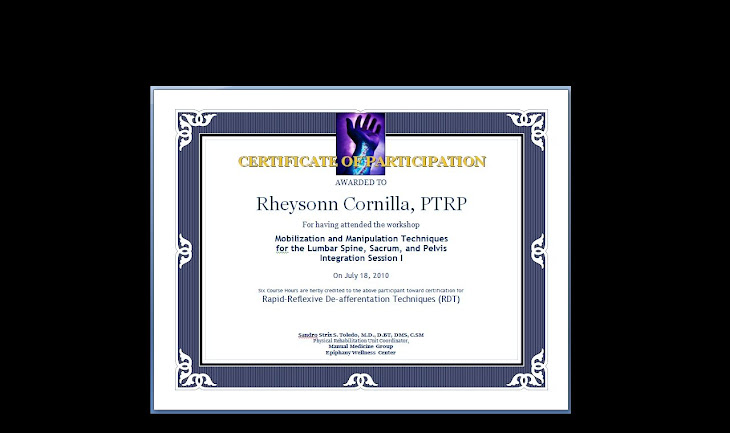
Reflexive De-afferentation Techniques (RDTs) have evolved from Neuro-myofascial techniques. Pain cycles easily broken with NMF techniques include headaches, nape and shoulder pain, TMJ pain, knee and foot pain. Mechanical and repetitive strain injuries that may be addressed with NMF-T include mechanical low back pain syndromes, thoracic outlet syndromes, “frozen shoulder,” carpal tunnel syndrome, lateral epicondylitis, and plantar fasciitis.
Articulatory techniques (ART)
Soft tissue injury and cumulative joint trauma result in joint ROM restrictions and / or pain. These techniques involve taking joints through their restrictive and compliant motion cycles. They improve joint range or motion and help ablate joint pain cycles.
Visceral Manipulation (VM, Ventral OMT techniques)
Trauma, lack of exercise, physical deconditioning, cumulative soft tissue injury, inflammation, surgery, or obesity can displace, restrict, tighten, or scar ligament and fascia that surround, support, or suspend organs. Fascial and ligamentous injury can result in focal strains that cause tissue or organ dysfunction and injury. Pain and inflammation cycles are facilitated. VM uses soft vectoral techniques aimed at re-engaging normal suspension or movement of internal tissues / organs.
The notion of tissue restrictions or compliance to movement extends to the viscera. Organs, fascia, mesentery, and visceral ligaments move along and in relation to each other in much the same way that we know normal fascia-muscle compartments do. This notion of mobility in relation to each other is differentiated from intrinsic organ motility. Organs are mobile in relation to each other and often possess an internal movement or motility. As all fascia moves with breathing, the lungs move as we breathe, the stomach and intestines move in relation to each other during peristalsis, the urinary bladder shifts location somewhat when we piss. Viscera also move around a bit with ambulation. Visceral mobility restrictions are thought to restrict normal fluid, solid, and impulse conduction through individual organs. VM addresses these restrictions.
Musculoskeletal conditions linked to visceral involvement include:
1. Mid to low back pain: The colon’s attachment to the back of the abdominal compartment may be strained. This can lend ligamentous tightness or displacement which, in turn, can inhibit normal spinal movement. These spinal segmental dysfunctions result in their related pain constellations.
2. Neck stiffness and pain: The suspensory ligaments of the lung and the pericardium all attach to cervical vertebrae. Ligamentous involvement here can cause cervical joint compression and flexion cervical somatic dysfunction. A forward head posture may thus actually be more closely related to a lung problem than to a thoracic spine dysfunction.
3. Shoulder restrictions and pain: Most paramedical personnel are familiar with the fact that gallbladder inflammation can result in pain that radiates to the right shoulder. Nerves that supply the suspensory ligaments of the liver stem from the spinal segment that innervates the shoulder. Liver suspensory ligament involvement may thus result in shoulder somatic dysfunctions.
4. Peripheral joint pain: Visceral compartment restrictions can transmit tension via adjacent myofascial trains into the limbs. These tension lines can cause compressive and torsional joint irritation and dysfunction.
5. Comparative Studies have found Visceral Manipulation also Beneficial for:
Somatic-Visceral Interactions
Chronic Spinal Dysfunction
Headaches and Migraine headaches
Carpal Tunnel Syndrome
Hip and Knee Pain
Sciatica
Whiplash
Seatbelt Injuries
Chest or Abdominal Sports Injuries
Visceral Manipulation has been known to improve:
1. Fatigue: Abnormal fascial tension or scarring from at the visceral suspension system may result from inflammation, surgery, and blunt or countercoup trauma. Loss of organ mobility due to restrictions of surrounding and supporting tissues have been linked to general fatigue. By example, lessened fatigue has been reported by whiplash-injured VA patients. Faster recovery rates and lower incidences of depressive symptoms have likewise been reported.
2. GI motility disorders: Ileus, constipation, and irritable bowel syndrome: Abnormal fascial tension or scarring from at the small and large intestinal suspension system may result from inflammation, surgery, or blunt trauma. This causes restrictions that negatively alter bowel motility and function.
3. Hemorrhoids: The venous drainage from the rectum toward the liver is impeded when the hepatoduodenal ligament is abnormally tight. The resultant vascular congestion can cause or aggravate hemorrhoids.
4. Incontinence: Ligamentous tightness and muscle hypertonus may limit proper urinary bladder expansion and emptying.
5. Comparative Studies have found Visceral Manipulation Beneficial for:
Digestive Disorders
Bloating and Constipation
Nausea and Acid Reflux
GERD
Swallowing Dysfunctions
Women’s and Men’s Health Issues
Chronic Pelvic Pain
Endometriosis
Fibroids and Cysts
Dysmenorrhea
Bladder Incontinence
Prostate Dysfunction
Referred Testicular Pain
Effects of Menopause Pain Related to
Post-operative Scar Tissue
Post-infection Scar Tissue
Autonomic Mechanisms
Pediatric Issues
Constipation and Gastritis
Persistent Vomiting
Vesicoureteral Reflux
Infant Colic
Emotional Issues
Anxiety and Depression
Post-Traumatic Stress Disorder
We are at www.manualmed.bravehost.com www.manualmed.blogspot.com
Wednesday, January 14, 2009


Conversations with Colleagues: (Steve Myles, American D.O)
I've decided do start including communicae with colleagues as posts on this site. The intent is that fellow manual medicine practitioners here may glean inpiration and insight from the strenths of years of experience that our colleagues have gained.
STRIX:
I have a small rehab team here. We train our staff and pass on anything we gain.
Physical therapy as a profession here is rather.. poorly updated. It is only beginning to pick up on manual med / manipulative skills. That puts my team at a fair leading edge insofar as the functional gains they can deliver for our patients, but leaves them with a hunger to pick up anything they can from anywhere they can. The way things look, we are likely to go in and out the country as frequently and as long as necessary in the next few years to pick up the skillsets for manip. As we find even good orthopedic manip protocols to be often less remarkable than OMM / OMT, we hope to learn from as many osteopaths as we can. We are looking for a synthesis, or at least what works best. Thus far, All manual medicine looks similar in principles and practice somewhere down the line. Our biggest bottomline is still to help our patients as best we can. It's still sadly fairly common for patients here to waste 18 sessions to go nowhere with the predominant PT practice here. The reason? It's modality based. Almost fully. That's the second thing we hope to do here - to push for an evolution in manual practice.
STEVE:
I empathize with you. In osteopathy, we learn first to analyze the strain pattern and decide where and why to begin. Then, we may do the correction using whatever paradigm appears appropriate. Diagnosis first and foremost, the treatment choice is just the tool.
STRIX:
Most PTs and MDs here have even never heard of visceral manip. And there are sadly no DO institutions here.
My practice also includes primary care and occupational medicine. It is at heart integrative. I however do not have any homeopathic training as yet. We have medicare / TRICARE coverage here. Dozens of HMOs have birthed and died since 2000. HM insurance is neither standard nor required.
I hope this background makes sense enough for you to help us out with recommendations?
STEVE:
I am familiar with the plight of conscientious physicians and health practitioners working in a system that is more money and resource management than patient care.
I used to do courses (for about 20 yrs), taking health professionals in a few weekends from knowing nothing about osteopathic analysis to learning some principles to apply to any occasion. If you teach someone a technique and the patient's condition doesn't apply, the practitioner is lost. If you teach principles, then the practitioner can always figure out what to do and where to begin.
To start with homeopathy, you might consider the Homeopathic Course - I review about 20 remedies in an easy-to-learn format. After you have had success with this acute prescribing for about 6 months, you might contact me for other resources to expand your knowledge.
Both osteopathy and homeopathy are easily each lifetime studies. The amazing contributions you make to your patients health keep you enthusiastically studying and learning.
I seem to have started another book here, Strix. If we can get the time zones straight, perhaps we could set a time to talk on the phone.
Take care, Strix.
Steve
STRIX:
I have a small rehab team here. We train our staff and pass on anything we gain.
Physical therapy as a profession here is rather.. poorly updated. It is only beginning to pick up on manual med / manipulative skills. That puts my team at a fair leading edge insofar as the functional gains they can deliver for our patients, but leaves them with a hunger to pick up anything they can from anywhere they can. The way things look, we are likely to go in and out the country as frequently and as long as necessary in the next few years to pick up the skillsets for manip. As we find even good orthopedic manip protocols to be often less remarkable than OMM / OMT, we hope to learn from as many osteopaths as we can. We are looking for a synthesis, or at least what works best. Thus far, All manual medicine looks similar in principles and practice somewhere down the line. Our biggest bottomline is still to help our patients as best we can. It's still sadly fairly common for patients here to waste 18 sessions to go nowhere with the predominant PT practice here. The reason? It's modality based. Almost fully. That's the second thing we hope to do here - to push for an evolution in manual practice.
STEVE:
I empathize with you. In osteopathy, we learn first to analyze the strain pattern and decide where and why to begin. Then, we may do the correction using whatever paradigm appears appropriate. Diagnosis first and foremost, the treatment choice is just the tool.
STRIX:
Most PTs and MDs here have even never heard of visceral manip. And there are sadly no DO institutions here.
My practice also includes primary care and occupational medicine. It is at heart integrative. I however do not have any homeopathic training as yet. We have medicare / TRICARE coverage here. Dozens of HMOs have birthed and died since 2000. HM insurance is neither standard nor required.
I hope this background makes sense enough for you to help us out with recommendations?
STEVE:
I am familiar with the plight of conscientious physicians and health practitioners working in a system that is more money and resource management than patient care.
I used to do courses (for about 20 yrs), taking health professionals in a few weekends from knowing nothing about osteopathic analysis to learning some principles to apply to any occasion. If you teach someone a technique and the patient's condition doesn't apply, the practitioner is lost. If you teach principles, then the practitioner can always figure out what to do and where to begin.
To start with homeopathy, you might consider the Homeopathic Course - I review about 20 remedies in an easy-to-learn format. After you have had success with this acute prescribing for about 6 months, you might contact me for other resources to expand your knowledge.
Both osteopathy and homeopathy are easily each lifetime studies. The amazing contributions you make to your patients health keep you enthusiastically studying and learning.
I seem to have started another book here, Strix. If we can get the time zones straight, perhaps we could set a time to talk on the phone.
Take care, Strix.
Steve
Osteopathic Manipulative Treatment: Diagnosis, Treatment, and Results
Introduction
We say that pain from a specific segment, relayed via its dermatomal level, is “radicular” in origin.
In practice, sensorimotor deficits and reflex abnormalities may be noted when we have pain that radiates to an extremity. Segmental innervation dictates where to look for a neurogenic lesion.
Capsular, tendinous, and musculoskeletal pain are usually addressed conservatively, with anything from traditional modality-based physiotherapy, medication, and exercise prescription.
Special attention to palpatory and positional reactions of soft tissue enables us to identify somatic dysfunctions.
Diagnostics
Upon diagnosis, osteopaths establish deviations in the relative position of the different vertebrae by means of palpation and examination. To that end, an imaginary line is drawn between the fingers of the examining practitioner, which are placed on certain sites of the patient's body. These sites are determined on the spinal column, for example, by the spinous processes and the transverse processes, etc. Account is taken of the fact that the joint surfaces between the various vertebrae on the spinal column are practically all different in shape and size and their position relative to the co-ordinate system is consequently different each time. Knowledge of this is essential.
What are the deviations from the usual position that a vertebra in the co-ordinate system may display relative to the neighbouring caudally located segment? (The standard abbreviation used in osteopathic medicine is shown between brackets.)
1. The vertebra may rotate around the anterior-posterior axis (AP axis). It is assumed that this rotation continues so far, due for example to overloading, that it becomes locked in this position. This applies to cervical, thoracic and lumbar vertebrae. The vertebra tilts, as it were; it is in a lateroflexion position (LFP).
2. The vertebra may be displaced dorsally, both unilaterally and bilaterally, and will then be forced by the shape of the joint surfaces also to rotate slightly in the other two body axes. This applies to cervical, thoracic and lumbar vertebrae. The vertebra is displaced backwards, as it were, and we call this a dorsal displacement; it is in a dorsal position (DP).
3. The vertebra may shift sideways. Again, this is possible in the case of cervical, thoracic and lumbar vertebrae. Lumbar vertebrae present differently on palpation than cervical and thoracic vertebrae due to the shape of their joint surfaces. The vertebra shifts, as it were, laterally; it is in a lateral position (LP).
4. The vertebra may be displaced ventrocranially, either unilaterally or bilaterally. This is accompanied by rotation around a longitudinal axis and around the anterior-posterior axis; however, the latter occurs only in unilateral displacement. Bilateral displacements are seen after whiplash trauma: the vertebra is in a ventral position (VP).
5. The vertebra may rotate around a longitudinal axis. When this axis runs through the vertebral body, the dorsal part of the vertebra - in particular the spinous process - is displaced away from the median line. The vertebra rotates; this is a rotation position (RP). The positions may occur singly or in combination, i.e. a single vertebra could display all five of the deviations from the usual position at the same time.
How does one establish these deviations from the usual position in the separate parts of the spinal column?
In the case of the cervical vertebrae, the transverse processes and the spinous processes can be extremely well palpated, thereby allowing the position of the neck vertebrae to be well determined. The examination is done while the patient is seated with the head hanging down.
In the case of the thoracic vertebrae, the ribs give a magnified image of the position of the vertebrae. Together with palpation of the musculature adjacent to the spinous process on both sides while the patient is sitting straight and while the patient is lying in the ventral decubitus position, this provides sufficient indicators to determine the deviations from the usual position.
In the case of the lumbar vertebrae, the diagnosis is largely made while the patient is lying in the ventral decubitus position. The position of the individual spinous processes is examined and the position relative to the underlying vertebra is also determined, as well as the position of the palpating thumbs, pressed into the tissue lateral to the erector trunci muscle.
In addition, the position of the vertebrae relative to one another is also examined while the patient is lying in the left or right lateral decubitus position with the pelvis on a cushion. A properly functioning back should then show a certain curvature. The above-mentioned deviations from the usual position can soon be identified in this way.
The pelvis plays an important role in the diagnosis of deviations from the usual position in the spinal column. Three deviations may be established in the pelvis, viz.:
tilted sacrum relative to a vertical line;
distorted pelvis without fixation of the sacroiliac (SI) joints, and
distorted pelvis with fixation of one or two SI joints.
A tilted sacrum is established by placing the patient in the ventral decubitus position, if necessary with a cushion under the abdomen if the patient fails to relax sufficiently. You then stand at the patient's head and place both index fingers at left and right of the cranial end of the sacrum alongside the superior articular process and press the fingers in the caudal direction. When one finger then moves more caudally than the other, this indicates that the sacrum is tilted and clamped between the two halves of the pelvis. In many cases this can also be clearly seen when the patient is in the seated position. This deviation may be caused by an asymmetrical vertical force, as in:
falling on the buttocks;
sliding down the stairs while seated;
manual expression during labour.
A distorted pelvis is a pelvis in which one or both ossa are rotated into the SI joint in the rest position, causing one or both cristae iliaca to be more cranial than the other. This is observed with the patient seated by placing the fingers left and right purely lateral on the crests.
In the case of a distorted pelvis without fixation of the SI joints, the functioning of the SI joints is checked as follows: sit behind the (standing) patient and place the right thumb on the sacrum alongside the spina iliaca posterior superior at the left side. A notional line is drawn to the left trochanter major and the left thumb is placed 2 cm away from the right thumb on this line. The patient is then asked to draw the left knee up to the abdomen, if necessary with the assistance of the hands. During the last part of the knee-lifting motion, the left thumb must describe a semi-circular movement and finish above the right thumb. If this happens, the functioning is in order. This examination applies to the left SI joint. For the right SI joint the placement of the hands should be symmetrically reversed.
The cause of the distorted pelvis may be due to deviations from the usual position of the lower lumbar vertebrae and/or to functional deviations or deviations from the usual position of the hip joint. A fused symphysis, which as syndesmosis should nonetheless show some mobility, may also be the cause of a distorted pelvis.
When no movement is observed in the SI joints bilaterally during functional examination of these joints in a distorted pelvis, there is nearly always a fairly appreciable (apparent) lower limb length discrepancy. This discrepancy is not true because it is based on the fact that the examination to determine the height of the two crests is repeated in the standing position and not in the seated position. If it is found upon repetition of this examination in the seated position that a tilted pelvis is still present, there is therefore mention of an 'apparent' lower limb length discrepancy.
A distorted pelvis due to bilateral SI joint locking is caused by fixation of one os ilium ventrally, as a result of which the acetabulum moves caudally; the other iliac bone is then fixed dorsally, which causes the acetabulum to move more towards the cranium. When these fixations are loosened, most of the torsion overstress is usually relieved. When the fingers are subsequently placed on the crests, bilaterally on the erector trunci muscle with the patient seated, one finger is frequently still found to be higher than the other; in this case a tilted pelvis is involved, where one of the os ilii has been displaced cranially or caudally.
A long existing tilted pelvis is 'cushioned' by the spinal column by allowing the vertebrae to rotate around the longitudinal axis, alternating left and right dorsally, up to and including C5. These twisting moments form a fixed pattern with fixed turning points. These fixed patterns are called 'formulae'. One of these formulae can be seen by asking a person with an anatomically normal back to rest one foot on an approximately 2 cm high platform and then stand with the knees straightened. One of the formulae - of which there are four - will then be formed. After removing the platform everything is restored to normal. The formulae are therefore physiological adjustments that can turn into deviations if they become fixed because the tilted pelvis becomes permanent. After correction of the tilted pelvis the formulae do not disappear unless they are treated adequately. This treatment, by manipulation, has a fixed, systematic sequence. The formulae are corrected in five treatments.
The cause of the tilted pelvis is excessive loading of the sacrum with opposing pressure in one of the acetabula; this occurs in sideward strain when lifting a load or in a fall on one of the os ischii.
The deviations from the usual position of the individual vertebrae are largely covered by the formulae. All the additional deviations of the vertebrae cannot be separately assessed and treated until approximately three treatments have been completed.
Complaints and symptoms
The above-mentioned deviations from the usual position of the vertebrae, which are often associated with dysfunctioning, frequently cause 'referred' complaints. The complaints are situated in the dermatome, viscerotome or myotome. In this way, for example, it is possible that complaints of pain in the leg with no radicular compression symptomatology on physical examination are caused by low lumbar deviations in the manual therapy sense. This also applies, of course, to cervicobrachialgia and to dizziness and headaches of diverse origin. With regard to the viscerotomes, it is noted that a connection is frequently found between diffuse cardiac complaints and deviations of the midthoracic spine. In nonspecific abdominal complaints a connection is also frequently seen with deviations of the lumbar spinal column. There is consequently also mention of a therapeutic approach to vegetative complaints that cannot be determined by means of instrumental diagnosis and which are susceptible to spinal column manipulation.
Therapy
Treatment consists generally of light pressure in the opposite direction to the deviation from the usual position, causing the vertebra to resume its normal position in the co-ordinate system. This will usually require several combined manipulations. It has been demonstrated empirically that the spinal column is governed by fixed laws. These laws entail, among other things, that:
each vertebra must be treated separately;
each deviation from the usual position must be treated separately, and
there is a certain sequence in the treatment of these deviations from the usual position.
This local and specific pressure is exerted on the transverse process or spinous process, in the course of which the adjacent segments either have to be relaxed or possibly fixed in torsion. During this treatment, use is made of cushions of different heights.
For reprints and communicae, please contact M. SICKESZ AND E.B. BONGARTZ. The authors would like to express their thanks to their colleague E. Keijzer, M.D., Doctor of Osteopathy, of Lelystad, for his valuable comments.
References
Cyriax F. Textbook of orthopaedic medicine. Vol. I. II. London: Baillière Tindall, 1980.
Lewit K. Manuele therapie. Part 1 and 2. Lochem: De Tijdstroom, 1979.
Niboyet JEN. La pratique de la médicine manuelle. Saint-Ruffine: Miasonneuve, 1968.
Williams PC. The lumbosacral spine. New York: McGraw-Hill, 1965.
Sickesz M. Orthomanipulatie. Alphen a.d. Rijn: Stafleu, 1981.
Biesinger E. Diagnosis and therapy of vertebrogenic vertigo. Laryngol Rhinol Otol (Stuttg) 1987; 66: 32-6.
Kunert W. Wirbelsäule, vegetatives Nervensystem und innere Medizin. Stuttgart: Enke Verlag, 1978.
We say that pain from a specific segment, relayed via its dermatomal level, is “radicular” in origin.
In practice, sensorimotor deficits and reflex abnormalities may be noted when we have pain that radiates to an extremity. Segmental innervation dictates where to look for a neurogenic lesion.
Capsular, tendinous, and musculoskeletal pain are usually addressed conservatively, with anything from traditional modality-based physiotherapy, medication, and exercise prescription.
Special attention to palpatory and positional reactions of soft tissue enables us to identify somatic dysfunctions.
Diagnostics
Upon diagnosis, osteopaths establish deviations in the relative position of the different vertebrae by means of palpation and examination. To that end, an imaginary line is drawn between the fingers of the examining practitioner, which are placed on certain sites of the patient's body. These sites are determined on the spinal column, for example, by the spinous processes and the transverse processes, etc. Account is taken of the fact that the joint surfaces between the various vertebrae on the spinal column are practically all different in shape and size and their position relative to the co-ordinate system is consequently different each time. Knowledge of this is essential.
What are the deviations from the usual position that a vertebra in the co-ordinate system may display relative to the neighbouring caudally located segment? (The standard abbreviation used in osteopathic medicine is shown between brackets.)
1. The vertebra may rotate around the anterior-posterior axis (AP axis). It is assumed that this rotation continues so far, due for example to overloading, that it becomes locked in this position. This applies to cervical, thoracic and lumbar vertebrae. The vertebra tilts, as it were; it is in a lateroflexion position (LFP).
2. The vertebra may be displaced dorsally, both unilaterally and bilaterally, and will then be forced by the shape of the joint surfaces also to rotate slightly in the other two body axes. This applies to cervical, thoracic and lumbar vertebrae. The vertebra is displaced backwards, as it were, and we call this a dorsal displacement; it is in a dorsal position (DP).
3. The vertebra may shift sideways. Again, this is possible in the case of cervical, thoracic and lumbar vertebrae. Lumbar vertebrae present differently on palpation than cervical and thoracic vertebrae due to the shape of their joint surfaces. The vertebra shifts, as it were, laterally; it is in a lateral position (LP).
4. The vertebra may be displaced ventrocranially, either unilaterally or bilaterally. This is accompanied by rotation around a longitudinal axis and around the anterior-posterior axis; however, the latter occurs only in unilateral displacement. Bilateral displacements are seen after whiplash trauma: the vertebra is in a ventral position (VP).
5. The vertebra may rotate around a longitudinal axis. When this axis runs through the vertebral body, the dorsal part of the vertebra - in particular the spinous process - is displaced away from the median line. The vertebra rotates; this is a rotation position (RP). The positions may occur singly or in combination, i.e. a single vertebra could display all five of the deviations from the usual position at the same time.
How does one establish these deviations from the usual position in the separate parts of the spinal column?
In the case of the cervical vertebrae, the transverse processes and the spinous processes can be extremely well palpated, thereby allowing the position of the neck vertebrae to be well determined. The examination is done while the patient is seated with the head hanging down.
In the case of the thoracic vertebrae, the ribs give a magnified image of the position of the vertebrae. Together with palpation of the musculature adjacent to the spinous process on both sides while the patient is sitting straight and while the patient is lying in the ventral decubitus position, this provides sufficient indicators to determine the deviations from the usual position.
In the case of the lumbar vertebrae, the diagnosis is largely made while the patient is lying in the ventral decubitus position. The position of the individual spinous processes is examined and the position relative to the underlying vertebra is also determined, as well as the position of the palpating thumbs, pressed into the tissue lateral to the erector trunci muscle.
In addition, the position of the vertebrae relative to one another is also examined while the patient is lying in the left or right lateral decubitus position with the pelvis on a cushion. A properly functioning back should then show a certain curvature. The above-mentioned deviations from the usual position can soon be identified in this way.
The pelvis plays an important role in the diagnosis of deviations from the usual position in the spinal column. Three deviations may be established in the pelvis, viz.:
tilted sacrum relative to a vertical line;
distorted pelvis without fixation of the sacroiliac (SI) joints, and
distorted pelvis with fixation of one or two SI joints.
A tilted sacrum is established by placing the patient in the ventral decubitus position, if necessary with a cushion under the abdomen if the patient fails to relax sufficiently. You then stand at the patient's head and place both index fingers at left and right of the cranial end of the sacrum alongside the superior articular process and press the fingers in the caudal direction. When one finger then moves more caudally than the other, this indicates that the sacrum is tilted and clamped between the two halves of the pelvis. In many cases this can also be clearly seen when the patient is in the seated position. This deviation may be caused by an asymmetrical vertical force, as in:
falling on the buttocks;
sliding down the stairs while seated;
manual expression during labour.
A distorted pelvis is a pelvis in which one or both ossa are rotated into the SI joint in the rest position, causing one or both cristae iliaca to be more cranial than the other. This is observed with the patient seated by placing the fingers left and right purely lateral on the crests.
In the case of a distorted pelvis without fixation of the SI joints, the functioning of the SI joints is checked as follows: sit behind the (standing) patient and place the right thumb on the sacrum alongside the spina iliaca posterior superior at the left side. A notional line is drawn to the left trochanter major and the left thumb is placed 2 cm away from the right thumb on this line. The patient is then asked to draw the left knee up to the abdomen, if necessary with the assistance of the hands. During the last part of the knee-lifting motion, the left thumb must describe a semi-circular movement and finish above the right thumb. If this happens, the functioning is in order. This examination applies to the left SI joint. For the right SI joint the placement of the hands should be symmetrically reversed.
The cause of the distorted pelvis may be due to deviations from the usual position of the lower lumbar vertebrae and/or to functional deviations or deviations from the usual position of the hip joint. A fused symphysis, which as syndesmosis should nonetheless show some mobility, may also be the cause of a distorted pelvis.
When no movement is observed in the SI joints bilaterally during functional examination of these joints in a distorted pelvis, there is nearly always a fairly appreciable (apparent) lower limb length discrepancy. This discrepancy is not true because it is based on the fact that the examination to determine the height of the two crests is repeated in the standing position and not in the seated position. If it is found upon repetition of this examination in the seated position that a tilted pelvis is still present, there is therefore mention of an 'apparent' lower limb length discrepancy.
A distorted pelvis due to bilateral SI joint locking is caused by fixation of one os ilium ventrally, as a result of which the acetabulum moves caudally; the other iliac bone is then fixed dorsally, which causes the acetabulum to move more towards the cranium. When these fixations are loosened, most of the torsion overstress is usually relieved. When the fingers are subsequently placed on the crests, bilaterally on the erector trunci muscle with the patient seated, one finger is frequently still found to be higher than the other; in this case a tilted pelvis is involved, where one of the os ilii has been displaced cranially or caudally.
A long existing tilted pelvis is 'cushioned' by the spinal column by allowing the vertebrae to rotate around the longitudinal axis, alternating left and right dorsally, up to and including C5. These twisting moments form a fixed pattern with fixed turning points. These fixed patterns are called 'formulae'. One of these formulae can be seen by asking a person with an anatomically normal back to rest one foot on an approximately 2 cm high platform and then stand with the knees straightened. One of the formulae - of which there are four - will then be formed. After removing the platform everything is restored to normal. The formulae are therefore physiological adjustments that can turn into deviations if they become fixed because the tilted pelvis becomes permanent. After correction of the tilted pelvis the formulae do not disappear unless they are treated adequately. This treatment, by manipulation, has a fixed, systematic sequence. The formulae are corrected in five treatments.
The cause of the tilted pelvis is excessive loading of the sacrum with opposing pressure in one of the acetabula; this occurs in sideward strain when lifting a load or in a fall on one of the os ischii.
The deviations from the usual position of the individual vertebrae are largely covered by the formulae. All the additional deviations of the vertebrae cannot be separately assessed and treated until approximately three treatments have been completed.
Complaints and symptoms
The above-mentioned deviations from the usual position of the vertebrae, which are often associated with dysfunctioning, frequently cause 'referred' complaints. The complaints are situated in the dermatome, viscerotome or myotome. In this way, for example, it is possible that complaints of pain in the leg with no radicular compression symptomatology on physical examination are caused by low lumbar deviations in the manual therapy sense. This also applies, of course, to cervicobrachialgia and to dizziness and headaches of diverse origin. With regard to the viscerotomes, it is noted that a connection is frequently found between diffuse cardiac complaints and deviations of the midthoracic spine. In nonspecific abdominal complaints a connection is also frequently seen with deviations of the lumbar spinal column. There is consequently also mention of a therapeutic approach to vegetative complaints that cannot be determined by means of instrumental diagnosis and which are susceptible to spinal column manipulation.
Therapy
Treatment consists generally of light pressure in the opposite direction to the deviation from the usual position, causing the vertebra to resume its normal position in the co-ordinate system. This will usually require several combined manipulations. It has been demonstrated empirically that the spinal column is governed by fixed laws. These laws entail, among other things, that:
each vertebra must be treated separately;
each deviation from the usual position must be treated separately, and
there is a certain sequence in the treatment of these deviations from the usual position.
This local and specific pressure is exerted on the transverse process or spinous process, in the course of which the adjacent segments either have to be relaxed or possibly fixed in torsion. During this treatment, use is made of cushions of different heights.
For reprints and communicae, please contact M. SICKESZ AND E.B. BONGARTZ. The authors would like to express their thanks to their colleague E. Keijzer, M.D., Doctor of Osteopathy, of Lelystad, for his valuable comments.
References
Cyriax F. Textbook of orthopaedic medicine. Vol. I. II. London: Baillière Tindall, 1980.
Lewit K. Manuele therapie. Part 1 and 2. Lochem: De Tijdstroom, 1979.
Niboyet JEN. La pratique de la médicine manuelle. Saint-Ruffine: Miasonneuve, 1968.
Williams PC. The lumbosacral spine. New York: McGraw-Hill, 1965.
Sickesz M. Orthomanipulatie. Alphen a.d. Rijn: Stafleu, 1981.
Biesinger E. Diagnosis and therapy of vertebrogenic vertigo. Laryngol Rhinol Otol (Stuttg) 1987; 66: 32-6.
Kunert W. Wirbelsäule, vegetatives Nervensystem und innere Medizin. Stuttgart: Enke Verlag, 1978.
Subscribe to:
Posts (Atom)


 Posts
Posts

